A clinic visit begins with diagnosis, not an automatic laser or cautery appointment. You will learn how the clinician examines the lesion, selects a removal method, decides whether tissue needs histopathology, and explains the sensations, wound care and risks involved.
Key takeaways
- Expect diagnosis before freezing, cautery or excision.
- Tell the clinician about medicines, allergies and bleeding risks.
- Ask whether removed mole tissue will be sent for testing.
- Plan wound care and follow-up before leaving the clinic.
What the clinician checks before removing a wart or mole
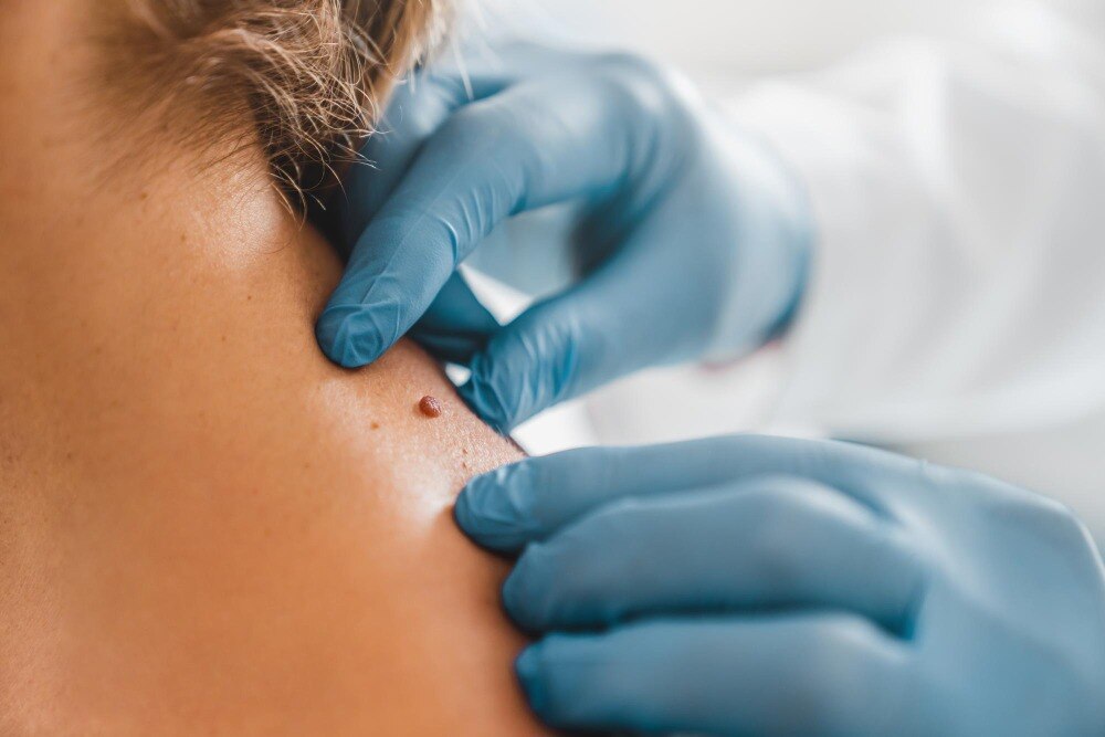
A wart removal consultation in Panvel begins with diagnosis, not automatic freezing or cautery. The clinician asks when the lesion appeared, whether it has changed, and whether it bleeds, itches or hurts. They review medicines, allergies, diabetes, immune problems, bleeding disorders and past keloid scars, then inspect its size, surface, colour, border and location.
Dermoscopy can reveal structures not visible to the naked eye.
A wart and mole removal process in Panvel changes direction when the lesion does not look typical.
- Asymmetry, an irregular border or several colours
- Recent enlargement or a new change in shape
- Spontaneous bleeding, persistent itching or a non-healing surface
- A dark, flat or atypical lesion mistaken for a wart
- Uncertainty between a wart, mole, skin tag, corn, callus, molluscum contagiosum, seborrhoeic keratosis or skin cancer
| Finding | Likely next step | Reason |
|---|---|---|
| Clearly typical wart | Routine wart treatment | The appearance and history support a confident diagnosis |
| Typical benign mole needing removal | Shave removal or planned excision | Technique depends on elevation, depth, site and desired tissue assessment |
| Suspicious mole | Biopsy or complete surgical excision | Tissue must be examined; destructive treatment could erase diagnostic evidence |
| Uncertain or complex lesion | Referral to a dermatologist or specialist | The diagnosis, margins or required pathology needs further assessment |
A biopsy removes a representative portion; excision removes the lesion, often with a margin and stitches. A clinician should explain scarring before consent, because even a small mole removal can leave a permanent mark.
What happens during the removal appointment
This is what happens during mole removal: the clinician confirms the site, reviews your consent, marks the lesion, and cleans the surrounding skin. Consent should cover the planned technique, possible pathology testing, bleeding, infection, recurrence and the chance of a permanent scar.
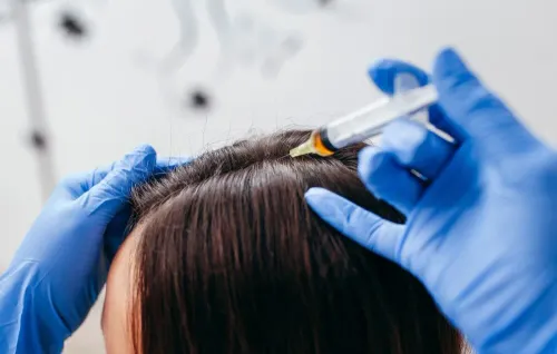
- Local anaesthetic is injected around the lesion. The needle can sting or burn for a few seconds; numbness follows.
- The clinician removes the wart or mole using the selected method. You should feel pressure, pulling or movement, not sharp pain. Tell them immediately if you feel pain.
- Bleeding is controlled with firm pressure, cautery or another method suited to the wound. A small ooze is common after treatment.
- A superficial wound may receive ointment and a small dressing. An excision that reaches deeper skin usually needs stitches and a more secure covering.
- The clinician explains when to remove or change the dressing, how to wash the area, and whether you need a follow-up visit.
Afterward, expect tenderness, tightness or mild throbbing once the anaesthetic wears off. Cryotherapy for a wart can produce cold, stinging and a blister; cautery can leave warmth or a burning sensation. Keep the site clean, avoid picking a scab or blister, and follow the clinic’s instructions for bathing and pain relief.
Contact the clinic for bleeding that will not stop with firm pressure, spreading redness, pus, fever or worsening pain.
Which removal method is used, and when is tissue tested?
The diagnosis determines the method, not the patient’s preferred technology. A clearly typical wart may be treated with liquid-nitrogen cryotherapy, electrocautery or radiofrequency ablation. A pigmented lesion should not be destroyed until the clinician is confident it is benign.
| Option | Best fit | Tissue testing |
|---|---|---|
| Cryotherapy | Typical superficial wart or selected benign lesion | No specimen remains |
| Electrocautery or radiofrequency | Small, raised benign lesions | Usually no specimen |
| Shave removal | Protruding, clinically benign mole | Send the tissue if diagnosis is uncertain |
| Punch biopsy or excision | Small lesion needing a deeper sample or complete removal | Histopathology is expected |
| Surgical excision | Suspicious, changing, or deeper mole; removes a margin | Histopathology is expected |
| Laser | Selected superficial cosmetic lesions after diagnosis | Not preferred when cancer must be excluded |
A raised, stable mole may suit shave removal, which leaves a flatter wound but can leave deeper cells behind. A flat lesion, or one with asymmetry, irregular borders, multiple colours, recent growth, bleeding, persistent itching or a non-healing surface, needs biopsy or complete excision rather than cosmetic destruction.
Microscopic histopathology decides whether wider treatment is necessary; appearance alone cannot do that.
If you are arranging a mole removal procedure Panvel appointment, request an assessment rather than booking laser by name. Ask whether the removed tissue will be labelled and sent to a pathology laboratory, and discuss the permanent-scar risk before consenting.
Why wart treatment follows a different pathway from mole removal
A wart treatment clinic in Panvel should confirm the lesion before destroying it. Warts result from human papillomavirus, while moles are melanocytic lesions; a pigmented or atypical growth must not receive wart treatment until the diagnosis is sufficiently clear.
An uncertain lesion may need dermoscopy, biopsy or excision because destruction can remove tissue needed for pathology.
| Treatment | What you may feel | What to expect |
|---|---|---|
| Cryotherapy for a confirmed wart | Brief cold pain, stinging, burning and later tenderness | A blister or crust can form; several sessions, often 2–3 weeks apart, may be needed |
| Electrocautery or ablation | Local-anaesthetic sting, followed by heat, pressure or a pulling sensation | Usually one treatment, but healing creates a small crust or wound |
| Mole removal | Anaesthetic sting, then pressure or pulling rather than sharp pain | Usually one planned procedure; tissue may be sent for pathology |
A wart can return because visible tissue removal does not guarantee immediate clearance of HPV from nearby skin. Keep the area clean, follow the dressing instructions, do not pick the crust or blister, and avoid shaving, biting or cutting across it. Do not share towels, nail tools or footwear used on an affected area.
Tell the clinician about diabetes, poor circulation, reduced sensation, immune suppression, pregnancy, blood thinners or abnormal scarring before treatment; cryotherapy and acids need extra caution on feet.
How to prepare for a Panvel consultation and plan recovery
Bring clear photographs of the lesion’s earlier appearance, previous biopsy or treatment reports, and a written list of medicines, supplements and allergies. Note when it appeared, whether it has grown, bled, itched or changed colour, and what you have already applied.
1. Disclose diabetes, poor circulation, reduced sensation, immune suppression, pregnancy or breastfeeding, and any history of keloid or unusually thick scars. These details affect wound healing and make cryotherapy or acid treatment risky, especially on the feet.
2. Name every blood thinner, including aspirin, clopidogrel, warfarin and apixaban. Do not stop one yourself; the clinician decides whether any change is safe.
3. Ask whether the clinician has examined the lesion with dermoscopy and confirmed a wart rather than a corn, callus, molluscum, seborrhoeic keratosis or skin cancer. A pigmented or changing lesion should not receive destructive wart treatment before diagnosis.
4. Confirm the proposed method, whether removed tissue will go for histopathology, whether stitches are needed, and when you can work, exercise, swim or expose the area to sunlight. Ask who handles a result that needs further treatment.
5. For a wart removal consultation Panvel patients should leave with wound-care instructions, pain advice and a review date. Dermaatriq can be assessed on these same practical points, not on the promise of a particular device.
If the proposed wart and mole removal process Panvel clinic outlines ignores diagnosis, pathology or your recovery constraints, seek another opinion.
Frequently asked questions
What does a clinician check before wart or mole removal?
The clinician asks when the lesion appeared and whether it changed, bled, itched or hurt. They also review medicines, allergies, diabetes, immune problems, bleeding disorders and previous keloid scars, then examine its size, surface, colour, border and location.
What happens during a wart or mole removal appointment?
The appointment begins with diagnosis and a skin examination. The clinician then discusses the suitable removal approach, explains what to expect, and provides instructions for caring for the treated area during recovery.
Which removal method is used for a wart or mole?
The method depends on the diagnosis, lesion features, location and clinical assessment. Wart treatment and mole removal follow different pathways, so the clinician selects the procedure after examination rather than automatically freezing or cauterising the lesion.
Is tissue tested after mole removal?
The clinician decides whether removed mole tissue should be sent for laboratory examination based on the lesion’s clinical features and diagnosis. Ask how the decision applies to your mole during the consultation.
How should I prepare for a Panvel wart or mole consultation?
Bring a list of medicines and allergies, mention diabetes or immune conditions, bleeding problems and past keloid scars, and describe when the lesion appeared and how it has changed. Ask about wound care, activity limits and follow-up before treatment.
Keywords